Background: The thymectomy specimens from the “thymectomy trial in non-thymomatous myasthenia gravis patients receiving prednisone therapy” (MGTX) [1] underwent rigid and comprehensive work-up, which results in a unique, spatially mapped dataset.
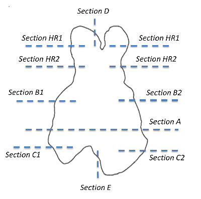
Fig. 1 Specimen work-up: Work-up scheme for the specimens in the MGTX trial [1, 2]. Thymuses were sub-divided into regions that underwent predefined evaluation (e.g. complete work-up of the central A-region, partial work-up of other regions)
Content: A xlsx-file with one table for patient data (“dataPatients” with age, gender etc.), one table with histomorphological data for the specimen regions (“dataAllRegions” with grading atrophy, grading overall fat etc.) and one table for the follicle morphology in the A region (“dataARegion” with n follicle with germinal centre etc.).
References: 1. Wolfe, G.I., et al., Randomized Trial of Thymectomy in Myasthenia Gravis. N Engl J Med, 2016. 375(6): p. 511-22. < /> 2. Ströbel, P., et al., The ageing and myasthenic thymus: A morphometric study validating a standard procedure in the histological workup of thymic specimens. Journal of Neuroimmunology, 2008. 201–202: p. 64-73. (2018-05-14)
